| |
Neuropathology
 |
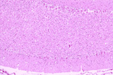
Section trough the cerebellum of an 8 week old Coton de TulÊar dog with early onset of rapidly progressive cerebellar ataxia.HE, 200X.

 What do you see? What do you see?
|
 |
 |
|  |
Answer: This is a massive depletion of the granule cells. Cerebellar granule cell atrophy is a neurodegenerative condition which has been described in a number of dogbreeds. In the Coton de TulÊar dog, the defect appears to be a congenital immunedefect leading to immunopathological destruction of the granule cells.
If you would like to learn more about veterinary neuropathology, visit the ESAVS course, 2007 in Bern Switzerland.
Course Masters in charge: Prof. Dr. Marc Vandevelde(CH)
Dr. Robert Higgins (USA), Dr. Georg Krinke (CH)
Overview:
This intensive course is specifically designed for diagnostic pathologists. We teach a systematic and practical approach to solve neuropathological problems starting from key morphologic features on macroscopical and histological examination. Further differentiation of such key features is trained by practical demonstration of a variety of specific lesions, which are than further studied in individual slide sessions. The course covers the major neurological diseases in domestic and laboratory animals. The course masters have taught this programme many times over the past several years. Course evaluations and feed back from former participants have shown that this concentrated effort is very effective and provides a solid base for further individual development in neuropathology.
Topics:
- General Neuropathology
Practical gross neuroanatomy for pathologists. Interpretation of the anamnesis, neurologic data. Braincutting/ representative sampling, processing. Gross lesions/ malformations. Basic reactions of the CNS to damage. General strategy in neurohistology. Wet lab: braincutting and representative sampling. Histolab: basic reaction patterns
- Inflammation
Principles of neuroimmunology and inflammation in the CNS. Special diagnostic techniques. Differential diagnosis of inflammatory/infectious diseases of the CNS: viral bacterial, protozoal, fungal, parasitic. Histolab: morphologic features of major inflammatory diseases
- Tumors and Malacias
Tumors of the nervous system in domestic animals. Tumors of the CNS in laboratory rodents. Trauma and infarcts. Toxic-metabolic diseases. Histolab: Tumors and malacias.
- Degenerations
Central nervous system/ picking up subtle lesions. Major types of degenerative lesions. Peripheral nervous system/muscle. Histolab: Degenerations.
- Spongy state in the CNS
Spongiform encephalopathies : BSE and Scrapie, an update. Other diagnostic methods for spongiform encephalopathies. Other spongy states/vacuoles in the CNS. Histolab: Spongiform encephalpathies and differential diagnosis
Download Registration Form
Tell a friend
|
Print version
|
Send this article
|
|  |

25th FECAVA EuroCongress 4-9 September 2019, St. Petersburg / RussiaESVN-ECVN Symposium 2018ESAVSVetAgendaLab in Practice - Clinical PathologyEuropean Master of Small Animal Veterinary MedicineSEVC 2014ESAVS - Neuropathology & MRICongressMed 2014ACVIM 2014VetContact
|


